


![[KO Validated] HADHA Rabbit mAb (A23892)](/upload/resize_cache/iblock/ddb/340_340_140cd750bba9870f18aada2478b24840a/2yg5rltzrzmgts5jvlf1wsi6ykdqcu8u.jpg "[KO Validated] HADHA Rabbit mAb (A23892)")
быстрая доставка ~4 недели
Размер
- 100 μL
- 200 μL
- 50 μL
[KO Validated] HADHA Rabbit mAb (A23892)
- Описание
- Характеристики
-
Overview
Product name [KO Validated] HADHA Rabbit mAb Catalog No. A23892 Host species Rabbit Purification method Affinity purification Isotype IgG CloneNo. ARC61206 Background
This gene encodes the alpha subunit of the mitochondrial trifunctional protein, which catalyzes the last three steps of mitochondrial beta-oxidation of long chain fatty acids. The mitochondrial membrane-bound heterocomplex is composed of four alpha and four beta subunits, with the alpha subunit catalyzing the 3-hydroxyacyl-CoA dehydrogenase and enoyl-CoA hydratase activities. Mutations in this gene result in trifunctional protein deficiency or LCHAD deficiency. The genes of the alpha and beta subunits of the mitochondrial trifunctional protein are located adjacent to each other in the human genome in a head-to-head orientation.Immunogen information
Immunogen Recombinant fusion protein containing a sequence corresponding to amino acids 545-763 of human HADHA (NP_000173.2). Sequence FYTTRCLAPMMSEVIRILQEGVDPKKLDSLTTSFGFPVGAATLVDEVGVDVAKHVAEDLGKVFGERFGGGNPELLTQMVSKGFLGRKSGKGFYIYQEGVKRKDLNSDMDSILASLKLPPKSEVSSDEDIQFRLVTRFVNEAVMCLQEGILATPAEGDIGAVFGLGFPPCLGGPFRFVDLYGAQKIVDRLKKYEAAYGKQFTPCQLLADHANSPNKKFYQ Gene ID Swiss Prot Synonyms GBP; ECHA; HADH; LCEH; MTPA; LCHAD; TP-ALPHA Calculated MW 83kDa Observed MW 75kDa Applications
Reactivity Human, Mouse, Rat Tested applications WB IF/ICC Recommended dilution - WB 1:1000-1:5000
- IF 1:50 - 1:200
Storage buffer Store at -20℃. Avoid freeze / thaw cycles.
Buffer: PBS with 0.05% proclin300, 0.05% BSA, 50% glycerol, pH7.3.Key application Western blotting Immunofluorescence Positive samples 293T, HepG2, NIH/3T3, Mouse kidney, Rat kidney, Rat liver Cellular location Mitochondrion 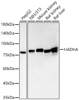
Western blot analysis of various lysates, using [KO Validated] HADHA Rabbit mAb (A23892) at 1:1000 dilution.
Secondary antibody: HRP Goat Anti-Rabbit IgG (H+L) (AS014) at 1:10000 dilution.
Lysates/proteins: 25ug per lane.
Blocking buffer: 3% nonfat dry milk in TBST.
Detection: ECL Basic Kit (RM00020).
Exposure time: 20s.
Western blot analysis of extracts from wild type(WT) and HADHA knockout (KO) 293T cells, using HADHA Rabbit mAb (A23892) at 1:1000 dilution.
Secondary antibody: HRP Goat Anti-Rabbit IgG (H+L) (AS014) at 1:10000 dilution.
Lysates/proteins: 25μg per lane.
Blocking buffer: 3% nonfat dry milk in TBST.
Detection: ECL Basic Kit (RM00020).
Exposure time: 20s.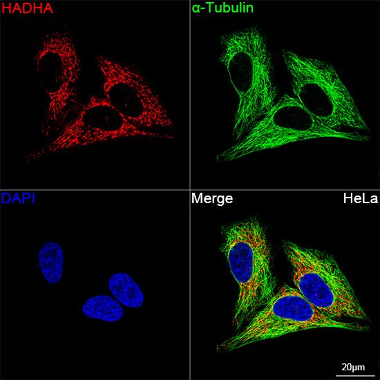
Confocal imaging of HeLa cells using HADHA Rabbit mAb (A23892, dilution 1:200)(Red). The cells were counterstained with α-Tubulin Mouse mAb (AC012, dilution 1:400) (Green). DAPI was used for nuclear staining (blue). Objective: 100x.
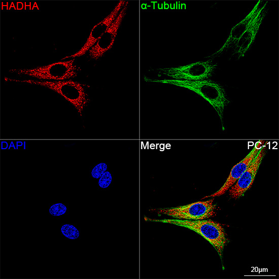
Confocal imaging of PC-12 cells using HADHA Rabbit mAb (A23892, dilution 1:200)(Red). The cells were counterstained with α-Tubulin Mouse mAb (AC012, dilution 1:400) (Green). DAPI was used for nuclear staining (blue). Objective: 100x.
-
Key applications Western blotting, Immunofluorescence Host species Кролик Antibody type Primary Antibodies Reactivity Человек, Мышь, Крыса Immunogen Recombinant fusion protein containing a sequence corresponding to amino acids 545-763 of human HADHA (NP_000173.2). KO Validated Validated Isotype IgG