


![[One Step] HSP70 Antibody Kit (RK05659)](/upload/resize_cache/iblock/d04/340_340_140cd750bba9870f18aada2478b24840a/wg4sjh2z35evoz4h2n7sypdk0pbfgpey.jpg "[One Step] HSP70 Antibody Kit (RK05659)")
быстрая доставка ~4 недели
Размер
- 50 μL
[One Step] HSP70 Antibody Kit (RK05659)
- Описание
- Характеристики
-
Overview
Product name [One Step] HSP70 Antibody Kit Catalog No. RK05659 Host species Purification method Isotype Background
Immunogen information
Immunogen Sequence Email for sequence Gene ID Swiss Prot Synonyms Calculated MW Observed MW Applications
Reactivity Tested applications Recommended dilution Storage buffer Key application Positive samples Cellular location 
Western blot analysis of extracts of HeLa cells,using GAPDH (AC001) antibody as the primary antibody at dilution of 1:80000.
Secondary antibody: using HRP Goat Anti-Rabbit IgG (H+L) antibody (AS014) at 1:4000-1:10000 dilution.
Lysates/proteins: 25μg per lane.
Blocking buffer: 3% nonfat dry milk in TBST.
Detection: ECL Basic Kit (RM00020).
Exposure time: 3s.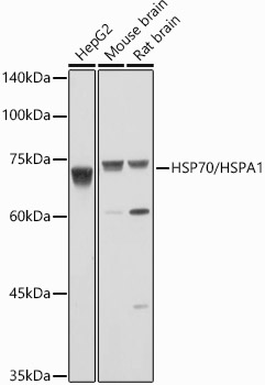
Western blot analysis of extracts of various cell lines, using HSP70/HSPA1 antibody (A0284) at 1:1000 dilution.
Secondary antibody: HRP Goat Anti-Rabbit IgG (H+L) (AS014) at 1:10000 dilution.
Lysates/proteins: 25μg per lane.
Blocking buffer: 3% nonfat dry milk in TBST.
Detection: ECL Basic Kit (RM00020).
Exposure time: 10s.
Immunohistochemistry analysis of paraffin-embedded rat kidney using HSP70/HSPA1 Rabbit pAb (A0284) at dilution of 1:100 (40x lens).Perform high pressure antigen retrieval with 10 mM citrate buffer pH 6.0 before commencing with IHC staining protocol.

Immunohistochemistry analysis of paraffin-embedded mouse spinal cord using HSP70/HSPA1 Rabbit pAb (A0284) at dilution of 1:100 (40x lens).Perform high pressure antigen retrieval with 10 mM citrate buffer pH 6.0 before commencing with IHC staining protocol.
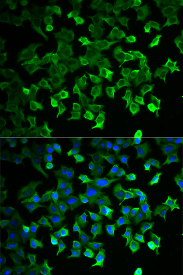
Immunofluorescence analysis of U2OS cells using HSP70/HSPA1 antibody (A0284). Blue: DAPI for nuclear staining.
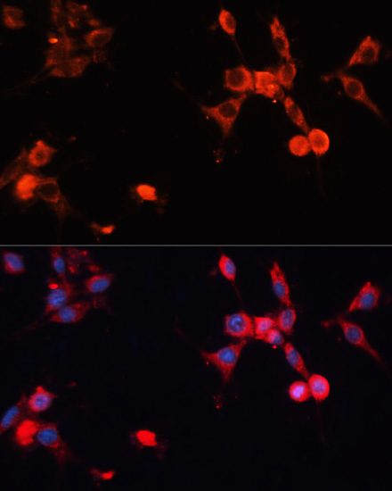
Immunofluorescence analysis of NIH/3T3 cells using HSP70/HSPA1 antibody (A0284) at dilution of 1:100. Blue: DAPI for nuclear staining.
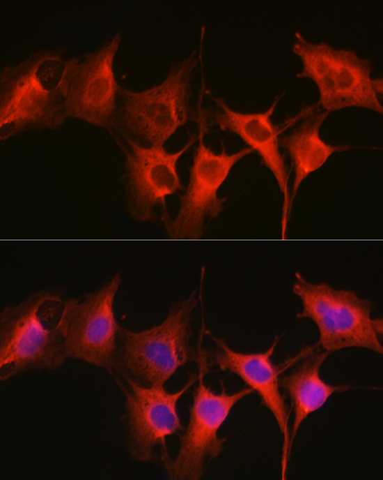
Immunofluorescence analysis of C6 cells using HSP70/HSPA1 Rabbit pAb (A0284) at dilution of 1:200 (40x lens). Blue: DAPI for nuclear staining.
-
Antibody type Primary Antibodies