


быстрая доставка ~4 недели
Размер
- 100 μL
- 20 μL
- 200 μL
- 50 μL
ABI1 Rabbit pAb (A16092)
- Описание
- Характеристики
-
Overview
Product name ABI1 Rabbit pAb Catalog No. A16092 Host species Rabbit Purification method Affinity purification Isotype IgG Background
This gene encodes a member of the Abelson-interactor family of adaptor proteins. These proteins facilitate signal transduction as components of several multiprotein complexes, and regulate actin polymerization and cytoskeletal remodeling through interactions with Abelson tyrosine kinases. The encoded protein plays a role in macropinocytosis as a component of the WAVE2 complex, and also forms a complex with EPS8 and SOS1 that mediates signal transduction from Ras to Rac. This gene may play a role in the progression of several malignancies including melanoma, colon cancer and breast cancer, and a t(10;11) chromosomal translocation involving this gene and the MLL gene has been associated with acute myeloid leukemia. Alternatively spliced transcript variants encoding multiple isoforms have been observed for this gene, and a pseudogene of this gene is located on the long arm of chromosome 14.Immunogen information
Immunogen Recombinant fusion protein containing a sequence corresponding to amino acids 180-230 of human ABI1 (NP_001012770). Sequence PMSGRGTLGRNTPYKTLEPVKPPTVPNDYMTSPARLGSQHSPGRTASLNQR Gene ID Swiss Prot Synonyms E3B1; ABI-1; ABLBP4; NAP1BP; SSH3BP; SSH3BP1 Calculated MW 55kDa Observed MW 55-65kDa Applications
Reactivity Human, Mouse, Rat Tested applications WB IHC-P Recommended dilution - WB 1:500 - 1:1000
- IHC-P 1:50 - 1:200
Storage buffer Store at -20℃. Avoid freeze / thaw cycles.
Buffer: PBS with 0.01% thimerosal, 50% glycerol, pH7.3.Key application Western blotting Immunohistochemistry Positive samples Neuro-2a, Rat brain Cellular location Cell junction, Cell projection, Cytoplasm, Nucleus, cytoskeleton, filopodium, growth cone, lamellipodium, postsynaptic cell membrane, postsynaptic density, synapse 
Western blot analysis of extracts of Rat brain cells, using ABI1 antibody (A16092) at 1:1000 dilution.
Secondary antibody: HRP Goat Anti-Rabbit IgG (H+L) (AS014) at 1:10000 dilution.
Lysates/proteins: 25μg per lane.
Blocking buffer: 3% nonfat dry milk in TBST.
Detection: ECL Basic Kit (RM00020).
Exposure time: 30s.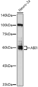
Western blot analysis of extracts of Neuro-2a cells, using ABI1 antibody (A16092) at 1:1000 dilution.
Secondary antibody: HRP Goat Anti-Rabbit IgG (H+L) (AS014) at 1:10000 dilution.
Lysates/proteins: 25μg per lane.
Blocking buffer: 3% nonfat dry milk in TBST.
Detection: ECL Enhanced Kit (RM00021).
Exposure time: 180s.
Immunohistochemistry analysis of paraffin-embedded rat ovary using ABI1 Rabbit pAb (A16092) at dilution of 1:50 (40x lens).Perform high pressure antigen retrieval with 10 mM citrate buffer pH 6.0 before commencing with IHC staining protocol.
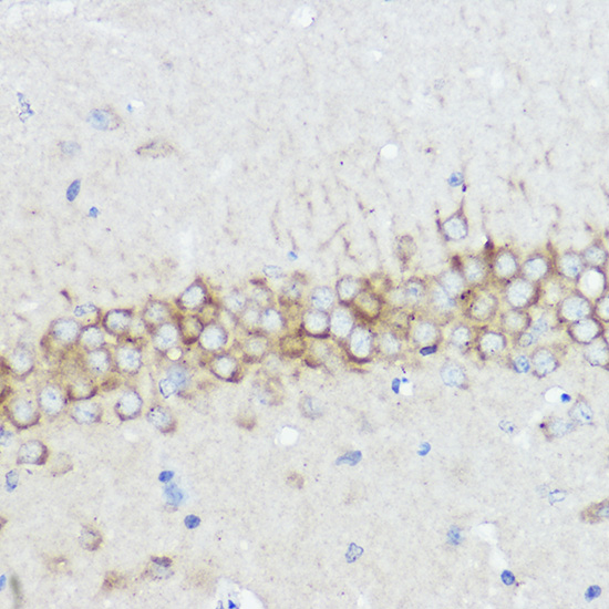
Immunohistochemistry analysis of paraffin-embedded rat brain using ABI1 Rabbit pAb (A16092) at dilution of 1:50 (40x lens).Perform high pressure antigen retrieval with 10 mM citrate buffer pH 6.0 before commencing with IHC staining protocol.
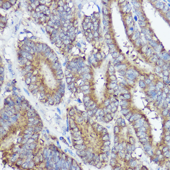
Immunohistochemistry analysis of paraffin-embedded human colon carcinoma using ABI1 Rabbit pAb (A16092) at dilution of 1:50 (40x lens).Perform high pressure antigen retrieval with 10 mM citrate buffer pH 6.0 before commencing with IHC staining protocol.
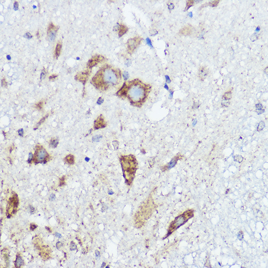
Immunohistochemistry analysis of paraffin-embedded mouse spinal cord using ABI1 Rabbit pAb (A16092) at dilution of 1:50 (40x lens).Perform high pressure antigen retrieval with 10 mM citrate buffer pH 6.0 before commencing with IHC staining protocol.
-
Key applications Western blotting, Immunohistochemistry Host species Кролик Antibody type Primary Antibodies Reactivity Человек, Мышь, Крыса Immunogen Recombinant fusion protein containing a sequence corresponding to amino acids 180-230 of human ABI1 (NP_001012770). Isotype IgG