


быстрая доставка ~4 недели
Размер
- 100 μL
- 200 μL
- 50 μL
α-Actinin-4 Rabbit mAb (A3379)
- Описание
- Характеристики
-
Overview
Product name α-Actinin-4 Rabbit mAb Catalog No. A3379 Host species Rabbit Purification method Affinity purification Isotype IgG CloneNo. ARC1959 Background
Alpha actinins belong to the spectrin gene superfamily which represents a diverse group of cytoskeletal proteins, including the alpha and beta spectrins and dystrophins. Alpha actinin is an actin-binding protein with multiple roles in different cell types. In nonmuscle cells, the cytoskeletal isoform is found along microfilament bundles and adherens-type junctions, where it is involved in binding actin to the membrane. In contrast, skeletal, cardiac, and smooth muscle isoforms are localized to the Z-disc and analogous dense bodies, where they help anchor the myofibrillar actin filaments. This gene encodes a nonmuscle, alpha actinin isoform which is concentrated in the cytoplasm, and thought to be involved in metastatic processes. Mutations in this gene have been associated with focal and segmental glomerulosclerosis.Immunogen information
Immunogen A synthetic peptide corresponding to a sequence within amino acids 1-100 of human α-Actinin-4 (O43707). Sequence MVDYHAANQSYQYGPSSAGNGAGGGGSMGDYMAQEDDWDRDLLLDPAWEKQQRKTFTAWCNSHLRKAGTQIENIDEDFRDGLKLMLLLEVISGERLPKPE Gene ID Swiss Prot Synonyms FSGS; FSGS1; ACTININ-4 Calculated MW 105kDa Observed MW 100kDa Applications
Reactivity Human, Mouse, Rat Tested applications WB IF/ICC IP Recommended dilution - WB 1:500 - 1:2000
- IF/ICC 1:50 - 1:200
- IP 1:50 - 1:200
Storage buffer Store at -20℃. Avoid freeze / thaw cycles.
Buffer: PBS with 0.02% sodium azide, 0.05% BSA, 50% glycerol, pH7.3.Key application Western blotting Immunofluorescence Immunoprecipitation Positive samples HeLa, 293T, Jurkat, Mouse brain, Mouse spleen, Rat lung Cellular location Cell junction, Cytoplasm, Nucleus Customer validation IF(Rattus norvegicus, Homo sapiens)

Western blot analysis of extracts of various cell lines, using α-Actinin-4 Rabbit mAb (A3379) at 1:1000 dilution.
Secondary antibody: HRP Goat Anti-Rabbit IgG (H+L) (AS014) at 1:10000 dilution.
Lysates/proteins: 25μg per lane.
Blocking buffer: 3% nonfat dry milk in TBST.
Detection: ECL Basic Kit (RM00020).
Exposure time: 30s.
Immunofluorescence analysis of C6 cells using α-Actinin-4 Rabbit mAb (A3379) at dilution of 1:100 (40x lens). Blue: DAPI for nuclear staining.
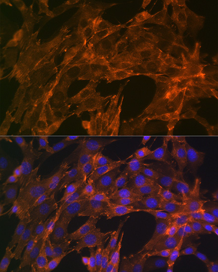
Immunofluorescence analysis of NIH-3T3 cells using α-Actinin-4 Rabbit mAb (A3379) at dilution of 1:100 (40x lens). Blue: DAPI for nuclear staining.

Immunofluorescence analysis of U-2 OS cells using α-Actinin-4 Rabbit mAb (A3379) at dilution of 1:100 (40x lens). Blue: DAPI for nuclear staining.

Immunoprecipitation analysis of 300 μg extracts of 293T cells using 3 μg α-Actinin-4 antibody (A3379). Western blot was performed from the immunoprecipitate using α-Actinin-4 antibody (A3379) at a dilution of 1:1000.
-
Key applications Western blotting, Immunofluorescence, Immunoprecipitation Host species Кролик Antibody type Primary Antibodies Reactivity Человек, Мышь, Крыса Immunogen A synthetic peptide corresponding to a sequence within amino acids 1-100 of human α-Actinin-4 (O43707). Isotype IgG