


![[KO Validated] SND1 Rabbit pAb (A5874)](/upload/resize_cache/iblock/e94/340_340_140cd750bba9870f18aada2478b24840a/uivn0pq47jp4ecage2oairz8fzk35u3i.jpg "[KO Validated] SND1 Rabbit pAb (A5874)")
быстрая доставка ~4 недели
Размер
- 100 μL
- 200 μL
- 50 μL
[KO Validated] SND1 Rabbit pAb (A5874)
- Описание
- Характеристики
-
Overview
Product name [KO Validated] SND1 Rabbit pAb Catalog No. A5874 Host species Rabbit Purification method Affinity purification Isotype IgG Background
This gene encodes a transcriptional co-activator that interacts with the acidic domain of Epstein-Barr virus nuclear antigen 2 (EBNA 2), a transcriptional activator that is required for B-lymphocyte transformation. Other transcription factors that interact with this protein are signal transducers and activators of transcription, STATs. This protein is also thought to be essential for normal cell growth. A similar protein in mammals and other organisms is a component of the RNA-induced silencing complex (RISC).Immunogen information
Immunogen Recombinant fusion protein containing a sequence corresponding to amino acids 26-285 of human SND1 (NP_055205.2). Sequence MVLSGCAIIVRGQPRGGPPPERQINLSNIRAGNLARRAAATQPDAKDTPDEPWAFPAREFLRKKLIGKEVCFTIENKTPQGREYGMIYLGKDTNGENIAESLVAEGLATRREGMRANNPEQNRLSECEEQAKAAKKGMWSEGNGSHTIRDLKYTIENPRHFVDSHHQKPVNAIIEHVRDGSVVRALLLPDYYLVTVMLSGIKCPTFRREADGSETPEPFAAEAKFFTESRLLQRDVQIILESCHNQNILGTILHPNGNIT Gene ID Swiss Prot Synonyms p100; TDRD11; Tudor-SN Calculated MW 102kDa Observed MW 120kDa Applications
Reactivity Human, Mouse, Rat Tested applications WB IHC-P IF/ICC IP Recommended dilution - WB 1:500 - 1:2000
- IHC-P 1:50 - 1:200
- IF/ICC 1:50 - 1:200
- IP 1:500 - 1:1000
Storage buffer Store at -20℃. Avoid freeze / thaw cycles.
Buffer: PBS with 0.01% thimerosal, 50% glycerol, pH7.3.Key application Western blotting Immunohistochemistry Immunofluorescence Immunoprecipitation Positive samples HeLa, Jurkat, HL-60, MCF7, 293T, Mouse liver, Rat liver Cellular location Cytoplasm, Melanosome, Nucleus Customer validation WB(Homo sapiens, Mus musculus)
Co-IP(Mus musculus)
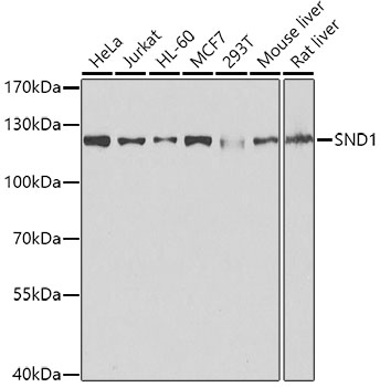
Western blot analysis of extracts of various cell lines, using SND1 antibody (A5874) at 1:1000 dilution.
Secondary antibody: HRP Goat Anti-Rabbit IgG (H+L) (AS014) at 1:10000 dilution.
Lysates/proteins: 25μg per lane.
Blocking buffer: 3% nonfat dry milk in TBST.
Detection: ECL Basic Kit (RM00020).
Exposure time: 1s.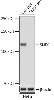
Western blot analysis of extracts from normal (control) and SND1 Rabbit pAb knockout (KO) HeLa cells, using SND1 Rabbit pAb antibody (A5874) at 1:3000 dilution.
Secondary antibody: HRP Goat Anti-Rabbit IgG (H+L) (AS014) at 1:10000 dilution.
Lysates/proteins: 25μg per lane.
Blocking buffer: 3% nonfat dry milk in TBST.
Detection: ECL Basic Kit (RM00020).
Exposure time: 1s.
Immunohistochemistry analysis of paraffin-embedded Mouse kidney using SND1 Rabbit pAb (A5874) at dilution of 1:100 (40x lens).Perform microwave antigen retrieval with 10 mM PBS buffer pH 7.2 before commencing with IHC staining protocol.
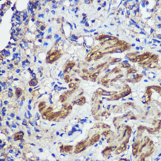
Immunohistochemistry analysis of paraffin-embedded Rat lung using SND1 Rabbit pAb (A5874) at dilution of 1:100 (40x lens).Perform microwave antigen retrieval with 10 mM PBS buffer pH 7.2 before commencing with IHC staining protocol.
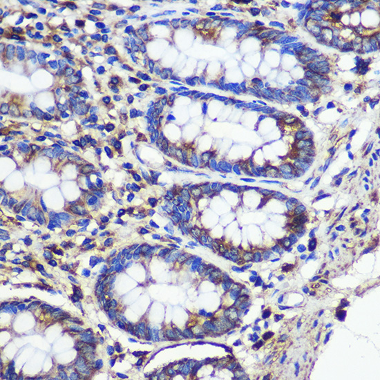
Immunohistochemistry analysis of paraffin-embedded Human colon using SND1 Rabbit pAb (A5874) at dilution of 1:100 (40x lens).Perform microwave antigen retrieval with 10 mM PBS buffer pH 7.2 before commencing with IHC staining protocol.

Immunofluorescence analysis of C6 cells using SND1 Rabbit pAb (A5874) at dilution of 1:100 (40x lens). Blue: DAPI for nuclear staining.
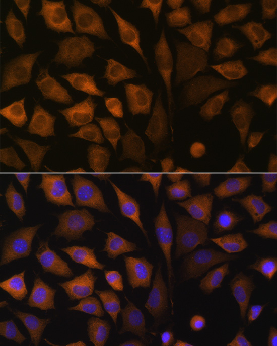
Immunofluorescence analysis of L929 cells using SND1 Rabbit pAb (A5874) at dilution of 1:100 (40x lens). Blue: DAPI for nuclear staining.
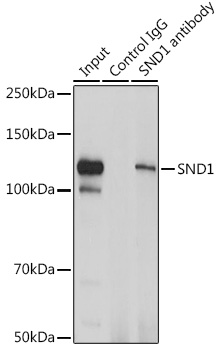
Immunoprecipitation analysis of 200 μg extracts of Jurkat cells using 3 μg SND1 antibody (A5874). Western blot was performed from the immunoprecipitate using SND1 antibody (A5874) at a dilution of 1:1000.
-
Key applications Western blotting, Immunohistochemistry, Immunofluorescence, Immunoprecipitation Host species Кролик Antibody type Primary Antibodies Reactivity Человек, Мышь, Крыса Immunogen Recombinant fusion protein containing a sequence corresponding to amino acids 26-285 of human SND1 (NP_055205.2). KO Validated Validated Isotype IgG