


![[KO Validated] Rap1A Rabbit pAb (A0975)](/upload/resize_cache/iblock/c1b/340_340_140cd750bba9870f18aada2478b24840a/i5adyh18t8m01kz319pkfe03ij04qvti.jpg "[KO Validated] Rap1A Rabbit pAb (A0975)")
быстрая доставка ~4 недели
Размер
- 100 μL
- 200 μL
- 50 μL
[KO Validated] Rap1A Rabbit pAb (A0975)
- Описание
- Характеристики
-
Overview
Product name [KO Validated] Rap1A Rabbit pAb Catalog No. A0975 Host species Rabbit Purification method Affinity purification Isotype IgG Background
This gene encodes a member of the Ras family of small GTPases. The encoded protein undergoes a change in conformational state and activity, depending on whether it is bound to GTP or GDP. This protein is activated by several types of guanine nucleotide exchange factors (GEFs), and inactivated by two groups of GTPase-activating proteins (GAPs). The activation status of the encoded protein is therefore affected by the balance of intracellular levels of GEFs and GAPs. The encoded protein regulates signaling pathways that affect cell proliferation and adhesion, and may play a role in tumor malignancy. Pseudogenes of this gene have been defined on chromosomes 14 and 17. Alternative splicing results in multiple transcript variants.Immunogen information
Immunogen A synthetic peptide corresponding to a sequence within amino acids 50-150 of human Rap1A (NP_002875.1). Sequence QCMLEILDTAGTEQFTAMRDLYMKNGQGFALVYSITAQSTFNDLQDLREQILRVKDTEDVPMILVGNKCDLEDERVVGKEQGQNLARQWCNCAFLESSAKS Gene ID Swiss Prot Synonyms RAP1; C21KG; G-22K; KREV1; KREV-1; SMGP21 Calculated MW 21kDa Observed MW 21kDa Applications
Reactivity Human, Mouse Tested applications WB IHC-P IF/ICC Recommended dilution - WB 1:500 - 1:1000
- IHC-P 1:50 - 1:100
- IF/ICC 1:50 - 1:100
Storage buffer Store at -20℃. Avoid freeze / thaw cycles.
Buffer: PBS with 0.01% thimerosal, 50% glycerol, pH7.3.Key application Western blotting Immunohistochemistry Immunofluorescence Positive samples 293T, U-87MG, HL-60 Cellular location Cell junction, Cell membrane, Cytoplasm, Early endosome, Lipid-anchor, perinuclear region Customer validation WB(Rattus norvegicus, Mus musculus)
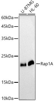
Western blot analysis of extracts of various cell lines, using Rap1A antibody (A0975) at 1:1000 dilution.
Secondary antibody: HRP Goat Anti-Rabbit IgG (H+L) (AS014) at 1:10000 dilution.
Lysates/proteins: 25μg per lane.
Blocking buffer: 3% nonfat dry milk in TBST.
Detection: ECL Basic Kit (RM00020).
Exposure time: 90s.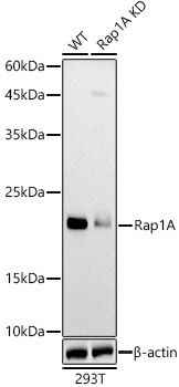
Western blot analysis of extracts from wild type(WT) and Rap1A knockdown (KD) 293T cells, using Rap1A antibody (A0975) at 1:1000 dilution.
Secondary antibody: HRP Goat Anti-Rabbit IgG (H+L) (AS014) at 1:10000 dilution.
Lysates/proteins: 25μg per lane.
Blocking buffer: 3% nonfat dry milk in TBST.
Detection: ECL Basic Kit (RM00020).
Exposure time: 90s.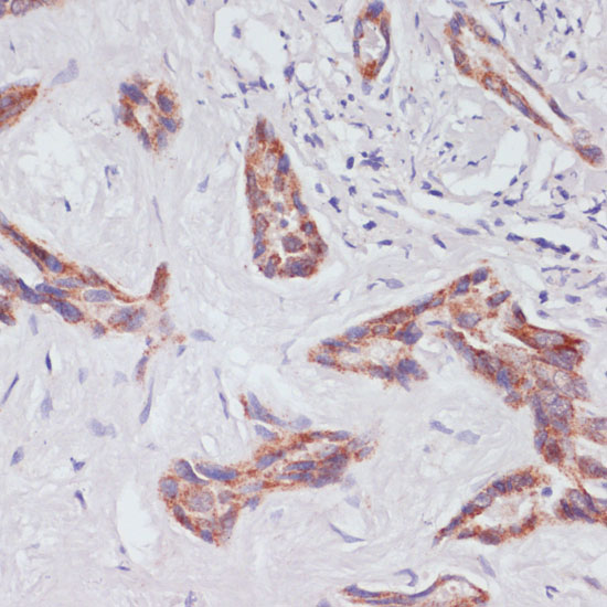
Immunohistochemistry analysis of paraffin-embedded human liver cancer using Rap1A antibody (A0975) at dilution of 1:100 (40x lens).Perform microwave antigen retrieval with 10 mM PBS buffer pH 7.2 before commencing with IHC staining protocol.

Immunohistochemistry analysis of paraffin-embedded human colon carcinoma using Rap1A antibody (A0975) at dilution of 1:100 (40x lens).Perform microwave antigen retrieval with 10 mM PBS buffer pH 7.2 before commencing with IHC staining protocol.
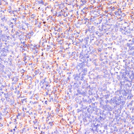
Immunohistochemistry analysis of paraffin-embedded mouse spleen using Rap1A antibody (A0975) at dilution of 1:100 (40x lens).Perform microwave antigen retrieval with 10 mM PBS buffer pH 7.2 before commencing with IHC staining protocol.
-
Key applications Western blotting, Immunohistochemistry, Immunofluorescence Host species Кролик Antibody type Primary Antibodies Reactivity Человек, Мышь Immunogen A synthetic peptide corresponding to a sequence within amino acids 50-150 of human Rap1A (NP_002875.1). KO Validated Validated Isotype IgG