


![[KD Validated] COX IV Rabbit pAb (A22871)](/upload/resize_cache/iblock/c8a/340_340_140cd750bba9870f18aada2478b24840a/lu91961nki08am08nefv59l05nr8u1kt.jpg "[KD Validated] COX IV Rabbit pAb (A22871)")
быстрая доставка ~4 недели
Размер
- 100 μL
- 200 μL
- 50 μL
[KD Validated] COX IV Rabbit pAb (A22871)
- Описание
- Характеристики
-
Overview
Product name [KD Validated] COX IV Rabbit pAb Catalog No. A22871 Host species Rabbit Purification method Affinity purification Isotype IgG Background
Cytochrome c oxidase (COX) is the terminal enzyme of the mitochondrial respiratory chain. It is a multi-subunit enzyme complex that couples the transfer of electrons from cytochrome c to molecular oxygen and contributes to a proton electrochemical gradient across the inner mitochondrial membrane. The complex consists of 13 mitochondrial- and nuclear-encoded subunits. The mitochondrially-encoded subunits perform the electron transfer and proton pumping activities. The functions of the nuclear-encoded subunits are unknown but they may play a role in the regulation and assembly of the complex. This gene encodes the nuclear-encoded subunit IV isoform 1 of the human mitochondrial respiratory chain enzyme. It is located at the 3' of the NOC4 (neighbor of COX4) gene in a head-to-head orientation, and shares a promoter with it. Pseudogenes related to this gene are located on chromosomes 13 and 14. Alternative splicing results in multiple transcript variants encoding different isoforms.Immunogen information
Immunogen Recombinant protein of human COX IV. Sequence Email for sequence Gene ID Swiss Prot Synonyms COX4; COXIV; COX4-1; COXIV-1; MC4DN16; COX IV-1 Calculated MW 20kDa Observed MW 17kDa Applications
Reactivity Human, Mouse, Rat Tested applications WB IHC-P IF/ICC Recommended dilution - WB 1:100 - 1:500
- IHC-P 1:50 - 1:200
- IF/ICC 1:50 - 1:200
Storage buffer Store at -20℃. Avoid freeze / thaw cycles.
Buffer: PBS with 0.01% thimerosal, 50% glycerol, pH7.3.Key application Western blotting Immunohistochemistry Immunofluorescence Positive samples 293T(KD), HepG2, Mouse heart, Rat heart Cellular location Mitochondrion inner membrane 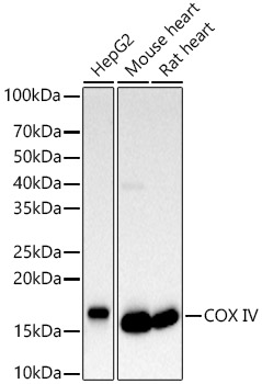
Western blot analysis of various lysates, using COX IV antibody (A22871) at 1:500 dilution.
Secondary antibody: HRP Goat Anti-Rabbit IgG (H+L) (AS014) at 1:10000 dilution.
Lysates/proteins: 25μg per lane.
Blocking buffer: 3% nonfat dry milk in TBST.
Detection: ECL Basic Kit (RM00020).
Exposure time: 10s.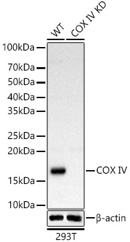
Western blot analysis of extracts from wild type(WT) and COX IVknockdown (KD) 293T(KD) cells, using COX IV antibody (A22871) at 1:500 dilution.
Secondary antibody: HRP Goat Anti-Rabbit IgG (H+L) (AS014) at 1:10000 dilution.
Lysates/proteins: 25μg per lane.
Blocking buffer: 3% nonfat dry milk in TBST.
Detection: ECL Basic Kit (RM00020).
Exposure time: 10s.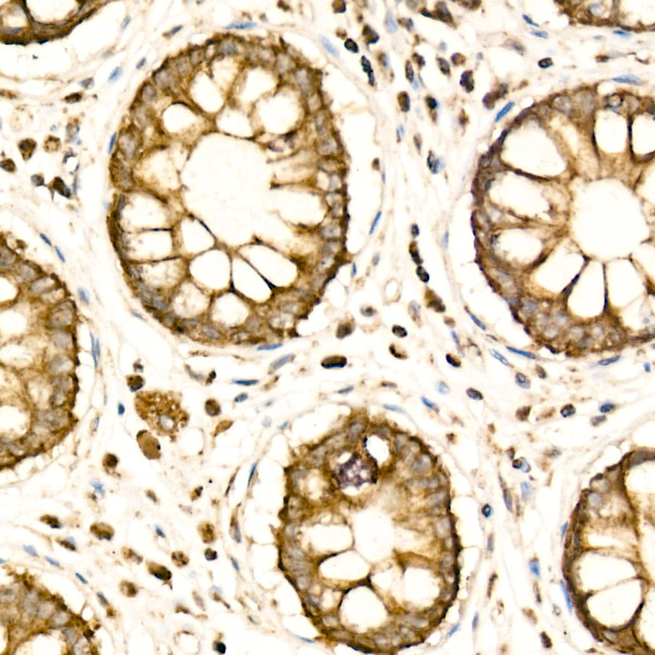
Immunohistochemistry analysis of paraffin-embedded human colon carcinoma using [KD Validated] COX IV Rabbit pAb (A22871) at dilution of 1:100 (40x lens).Perform high pressure antigen retrieval with 10 mM citrate buffer pH 6.0 before commencing with IHC staining protocol.
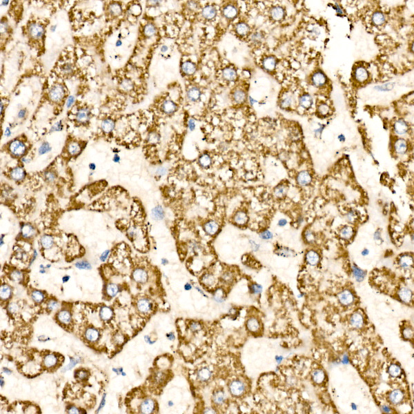
Immunohistochemistry analysis of paraffin-embedded human liver using [KD Validated] COX IV Rabbit pAb (A22871) at dilution of 1:100 (40x lens).Perform high pressure antigen retrieval with 10 mM citrate buffer pH 6.0 before commencing with IHC staining protocol.
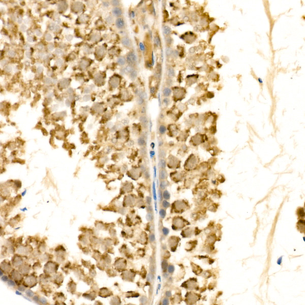
Immunohistochemistry analysis of paraffin-embedded mouse testis using [KD Validated] COX IV Rabbit pAb (A22871) at dilution of 1:100 (40x lens).Perform high pressure antigen retrieval with 10 mM citrate buffer pH 6.0 before commencing with IHC staining protocol.
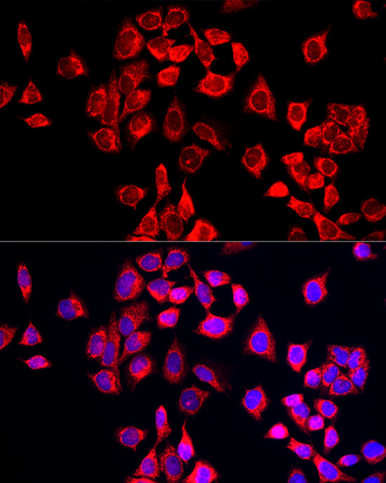
Immunofluorescence analysis of HeLa cells using [KD Validated] COX IV Rabbit pAb (A22871) at dilution of 1:50 (40x lens). Blue: DAPI for nuclear staining.

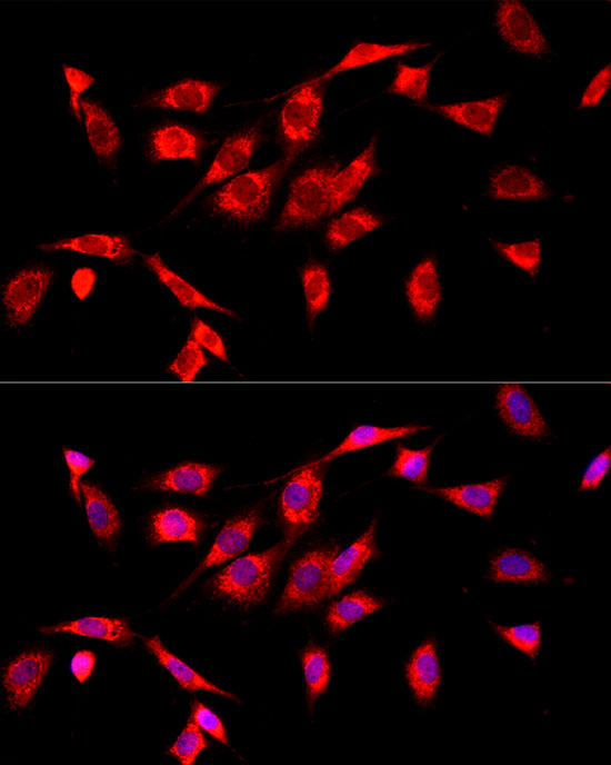
Immunofluorescence analysis of HepG2 cells using [KD Validated] COX IV Rabbit pAb (A22871) at dilution of 1:50 (40x lens). Blue: DAPI for nuclear staining.
Immunofluorescence analysis of NIH/3T3 cells using [KD Validated] COX IV Rabbit pAb (A22871) at dilution of 1:50 (40x lens). Blue: DAPI for nuclear staining.
-
Key applications Western blotting, Immunohistochemistry, Immunofluorescence Host species Кролик Antibody type Primary Antibodies Reactivity Человек, Мышь, Крыса Immunogen Recombinant protein of human COX IV. Isotype IgG